
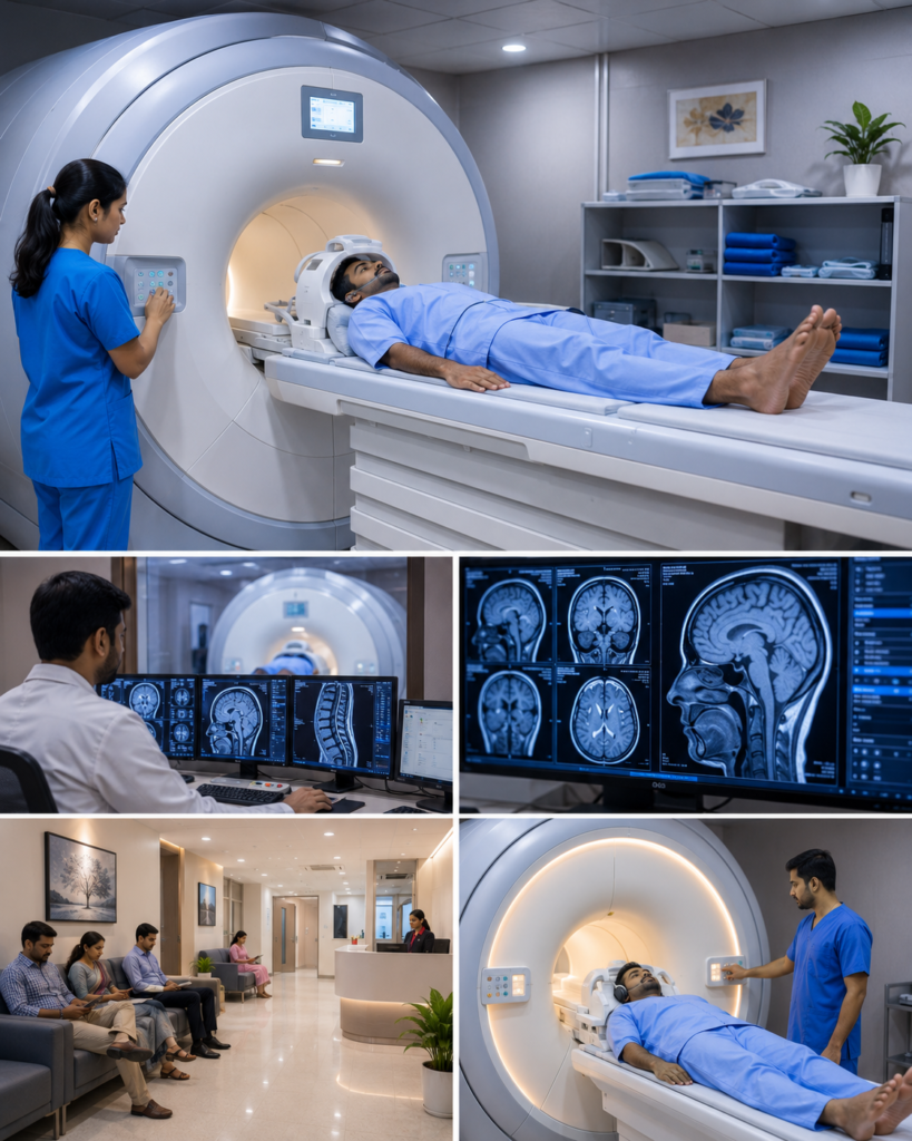
MRI Scan Centers in Madurai – A Complete Guide to Magnetic Resonance Imaging
Magnetic Resonance Imaging (MRI) is one of the most advanced diagnostic imaging techniques used to examine internal organs, soft tissues, joints, nerves, and the brain without using ionizing radiation. MRI scans provide highly detailed images that help doctors diagnose a wide range of medical conditions accurately and plan appropriate treatment.
If you’re searching for MRI Scan Centers in Madurai, this guide explains how MRI works, the conditions it helps diagnose, the types of MRI scans available, and how to prepare for the procedure. This article is intended for informational purposes and does not promote or recommend any specific MRI center.
What is an MRI Scan?
Magnetic Resonance Imaging (MRI) is a non-invasive imaging test that uses a powerful magnetic field, radio waves, and computer technology to create detailed images of organs, muscles, nerves, bones, and other internal structures.
Unlike X-rays and CT scans, MRI does not use ionizing radiation, making it an important imaging option for many medical conditions.
Why is an MRI Scan Performed?
Doctors may recommend an MRI scan to:
- Diagnose unexplained pain
- Evaluate brain disorders
- Detect spinal problems
- Assess joint injuries
- Examine muscles and ligaments
- Identify tumors
- Monitor treatment progress
- Detect internal organ abnormalities
- Evaluate blood vessels in selected cases
MRI is often used when detailed images of soft tissues are needed for diagnosis.
Common Types of MRI Scans
Brain MRI
A brain MRI may help evaluate:
- Stroke
- Brain tumors
- Multiple sclerosis
- Head injuries
- Chronic headaches
- Seizure disorders
- Brain infections
Spine MRI
Used to assess:
- Herniated discs
- Spinal stenosis
- Spinal cord disorders
- Nerve compression
- Degenerative spine disease
- Back pain
- Neck pain
Joint MRI
Commonly performed for:
- Knee injuries
- Shoulder injuries
- Hip disorders
- Ankle injuries
- Wrist problems
- Ligament tears
- Cartilage damage
Abdominal MRI
May be used to evaluate:
- Liver diseases
- Kidney disorders
- Pancreatic conditions
- Gallbladder abnormalities
- Adrenal gland disorders
Pelvic MRI
Used for assessing:
- Uterine conditions
- Ovarian disorders
- Prostate diseases
- Bladder abnormalities
- Pelvic tumors
Cardiac MRI
Provides detailed images of the heart and may help evaluate:
- Heart muscle disorders
- Congenital heart diseases
- Heart function
- Certain vascular abnormalities
Conditions Diagnosed with MRI
MRI is commonly used to investigate:
- Brain tumors
- Stroke
- Multiple sclerosis
- Spinal disc prolapse
- Arthritis
- Sports injuries
- Ligament tears
- Meniscus injuries
- Bone infections
- Soft tissue tumors
- Liver diseases
- Kidney abnormalities
- Pelvic disorders
- Certain heart conditions
The specific MRI examination depends on the patient’s symptoms and clinical evaluation.
MRI with Contrast
Some MRI examinations require a contrast agent to improve visualization of blood vessels, tumors, inflammation, or specific organs.
Your doctor will determine whether contrast is necessary based on your medical condition and the purpose of the scan.
Preparing for an MRI Scan
Preparation may include:
- Wearing comfortable clothing without metal fasteners
- Removing jewelry, watches, and metallic accessories
- Informing the healthcare team about implanted medical devices
- Discussing any history of kidney disease if contrast is planned
- Informing the radiology team if you are pregnant or think you may be pregnant
- Following fasting instructions if advised for specific examinations
Always follow the preparation instructions provided by your healthcare provider or imaging facility.
What Happens During the Procedure?
During the scan:
- You will lie on a movable examination table.
- The table slides into the MRI scanner.
- You must remain still while images are obtained.
- The machine produces loud tapping or knocking sounds during scanning.
- Communication with the radiology team is maintained throughout the examination.
The scan duration varies depending on the area being examined and whether contrast is used.
Benefits of MRI
MRI offers several advantages:
- No ionizing radiation
- Excellent soft tissue detail
- Non-invasive procedure
- High diagnostic accuracy
- Useful for complex neurological and musculoskeletal conditions
- Helps guide treatment planning
- Suitable for repeated follow-up imaging when clinically appropriate
Are There Any Risks?
MRI is generally considered safe for most individuals. However, certain implanted medical devices, metal fragments, or specific health conditions may require special precautions or alternative imaging methods.
Contrast-enhanced MRI may not be suitable for everyone, particularly individuals with certain kidney conditions. Your healthcare provider will assess whether the examination is appropriate for you.
Tips Before Your MRI Appointment
To help ensure a smooth examination:
- Arrive early for registration.
- Carry previous imaging reports if available.
- Inform the staff about pacemakers, implants, or previous surgeries.
- Mention any allergies or kidney problems if contrast may be used.
- Ask questions if you are concerned about claustrophobia or the procedure.
Frequently Asked Questions
Does an MRI scan use radiation?
No. MRI uses magnetic fields and radio waves rather than ionizing radiation.
How long does an MRI scan take?
The duration depends on the body part being examined and the type of study, but many MRI examinations are completed within 20 to 60 minutes.
Is an MRI scan painful?
No. MRI is a painless procedure. However, remaining still during the scan is important for obtaining clear images.
Can I eat before an MRI?
Many MRI scans do not require fasting, although certain examinations or contrast-enhanced studies may have specific preparation instructions.
Why is contrast used during an MRI?
Contrast material may improve visualization of certain organs, blood vessels, inflammation, or tumors, helping doctors obtain more detailed diagnostic information.
Conclusion
MRI is an advanced and highly effective diagnostic imaging technique that provides detailed views of the body’s soft tissues, organs, joints, brain, and spine without using ionizing radiation. It plays an important role in diagnosing neurological disorders, musculoskeletal injuries, abdominal diseases, and many other medical conditions. Understanding the purpose of an MRI scan, following the recommended preparation, and consulting a qualified healthcare professional can help ensure accurate diagnosis and appropriate treatment planning.